水浸対物レンズオプションを用いたハイコンテント共焦点イメージング
ImageXpress® Micro Confocalシステムは、固定細胞および生細胞のワイドフィールドイメージングと共焦点イメージングを切り替えできるハイコンテントイメージングソリューションです。生物全体、厚みのある組織、2Dおよび3Dモデル、細胞または細胞内の現象の高品質な画像を取得できます。スピニングディスク共焦点およびsCMOSカメラにより、速度を求められる心筋細胞の拍動や幹細胞分化などの特異的な現象もイメージングできます。MetaXpressソフトウェアと、水浸対物レンズなどの選択可能なフレキシブルなオプションを使用することで、3Dアッセイの開発からスクリーニングまで、多くの共焦点イメージングアプリケーションを利用できます。
ImageXpress Micro Confocal
ハイコンテントイメージングシステムの特長
-

高品質な画像を取得
特許取得済みのAgileOptix™スピニングディスク共焦点テクノロジーと、広い視野、明るい光源を利用して、コントラストに優れた高解像度の画像を取得できます。
-

画像取得と解析をカスタマイズ
画像取得および解析パラメーターを最大限制御することで、3D構造解析から生物または細胞集団内の特定のオブジェクトのターゲティングイメージングまで、様々なアプリケーションが利用できます。水浸対物レンズ、環境コントロールなどの標準オプションや、モレキュラーデバイスの高性能なカスタマイズソリューションを利用すれば、より多くのアプリケーションを実行できます。
-

短時間でより多くのデータを解析
MetaXpress® PowerCore™ソフトウェアはマルチコアCPU環境において画像処理作業を並列で処理し、ハイスループットを求められる環境での解析スピードをさらに高めます。
-
唯一無二のAgileOptixスピニングディスク共焦点テクノロジー
独自の光学系、高輝度固体光源および最新のsCMOSカメラにより、高い感度イメージングを提供します。共焦点光学系はクリックのみで高解像度モード、高速度モードに切り替えることが可能です。
-
一体型ロボティック・リキッドハンドリングオプション
化合物の添加、ウェルの洗浄、および培地交換を伴うアッセイのために、オプションの一体型ロボティック・リキッドハンドリングを利用できます。
-
正確な3D解析
3D解析モジュールは共焦点イメージングに最適化されており、体積と距離の3D測定が可能です。
-
複数のイメージングモード
標準で蛍光、ワイドフィールド、共焦点イメージングを搭載しています。オプションとして、位相差および明視野ラベルフリーイメージング、RGBカラーイメージング、水浸イメージングの観察法に対応しています。
*NEW* 標準オプションと高性能なカスタマイズオプション
によるイメージング機能の最適化
水浸対物レンズ、高輝度レーザー、生体深部組織に最適な低クロストーク透過共焦点ディスクモジュール、低酸素環境研究、厚みのある細胞サンプルおよびマルチプレックスアッセイなど様々なニーズに対応する最適化されたイメージング機能を提供しています。

詳細を見る
フレキシブルなイメージングソリューションを使って
研究を拡張できます
モレキュラーデバイスは、ImageXpress Micro Confocalハイコンテントイメージングシステム用に、フレキシブルなオプションを提供しています。ハンギングドロップ法や丸底・平底プレートなどの様々なサンプルフォーマットを用いた画像取得や、環境制御下での細胞のモニタリングなど、様々な研究ニーズを満たす柔軟性の高いシステムです。30年を超えるイメージングの経験がある当社が、アッセイに最適な画像を確実かつ正確なに取得することができるオプションの選択をお手伝いします。
 水浸対物レンズ
20X、40X、60Xの水浸対物レンズを用いると、画像取得時の光学収差精度が改善され、深さ方向の画像精度が向上し、短い露光時間でも明るい画像を取得できます。
 環境コントロールユニット
環境コントロールユニットを用いると、培地の蒸発を最低限に抑えながら温度と湿度を維持でき、複数日に渡る生細胞のタイムラプスイメージングが行えます。
 透過光ユニット
当社の光学位相差観察が可能な透過光ユニットと位相差対物レンズを用いると、非染色細胞でも容易に観察でき、バックグラウンドとも区別できる、コントラストの高い位相差画像を取得できます。
 リキッドハンドリングユニット
統合されたリキッドハンドリングユニットで、化合物の添加やウェルの洗浄、そして培地交換が必要なアッセイワークフローを自動化できます。
詳細を見る
水浸対物レンズ
水浸対物レンズは、シグナルを最高4倍高めることができます。短い露光時間で3次元(3D)で厚みのある組織サンプルをより深く観察する際に役立ちます。
迅速に観察を始めるための合理化されたセットアップ
水浸対物レンズはフィールドアップグレード可能で、現行のワークフローに最低限の変更を加えるだけで利用できます。

深い深度で最高4倍のシグナル強度
3Dサンプルをより正確に再構築するため、Z分解能を改善し、光学収差を低減しています。

鮮明ではっきりした画像品質
優れた集光効率により、短い露光時間でも明るい画像が得られ、データの品質向上に寄与します。

信頼性のある制御系統
“MetaXpressソフトウェアには、水浸制御システムの状態を示す統合された直感的なセンサーとアラートが含まれており、安心して水浸レンズを使用できます。”

ハイスループットな3Dサンプルの画像取得
水浸対物レンズを備えたImageXpress Micro Confocalシステムは、画像取得中、自動的に水を補充します。そのため、高いスループットを維持したままマイクロプレートサンプル全体の画像取得を行えます。

3D構造の表示と定量の単純化
Z平面のスタックとして取得されたスフェロイド、微小組織、3Dマトリックス中の細胞、および小さい生物を解析できます。MetaXpress 3D解析モジュールを用いて、体積、XYZ位置、隣接するオブジェクトとの距離、直径、深度、様々な輝度測定値、テクスチャー、またはオブジェクトの数を評価できます。

画像取得時間の短縮
QuickIDターゲット画像取得は、低倍率でイメージングを行い興味のあるオブジェクトや稀な現象を検出し、その後、必要な波長、Zスタック、または時点で自動的に高倍率画像を取得する機能です。 この機能により、選択した標的のみを高倍率で画像取得することで、画像取得時間を短縮し、画像の保存に必要な容量を減らすことができます。

役に立つソリューションを提供します
モレキュラーデバイスは、ImageXpress Micro
Confocalハイコンテントイメージングシステムをカスタマイズして、ユーザーのニーズに合わせた調整を行います。インキュベーター、リキッドハンドラー、フルオートメーションワークセル用のロボットなどの他社の装置とも統合できます。高品質の製品と世界的サポートの提供は、ライフサイエンス業界で30年を超える経験のある当社にお任せください。
販売は当社のCustom Product Purchase Terms(https://www.moleculardevices.com/custom-products-purchase-terms)に従って行います。
 高輝度レーザー
5または8チャンネル高輝度レーザーで実験能力を拡張できます。
 深部組織観察用共焦点ディスクモジュール
深部組織観察用 共焦点ディスクモジュールは、クロストークを低減し、焦点外の光ノイズを低減して、組織のより深い部分までクリアに観察可能にします。
ロボティックオートメーション
スループットを高め、ヒューマンエラーをなくし、無菌性を維持し、一貫したサンプル処理を行います。モジュールで構成要素を追加できる設計であり、アップグレード可能です。
 リアルタイム用量反応
1秒間に100フレーム超のリアルタイムストリーミングで画像取得しながら、同時に自動ピペッターで化合物を添加できます。
すぐに使えるハイスループット長期カイネティクス
温度、O2(低酸素)、CO2、および湿度を一定条件に保ちながら、複数のプレートを長期間にわたって保持しイメージングできます。生細胞観察できるプレート枚数を200プレート以上に拡張できます。
**
すぐに使えるハイスループットオルガノイドスクリーニングソリューションのためには培地交換が必要です。
高輝度レーザー
高出力レーザーによる励起により露光時間を最大75%減らせます†。5チャンネルレーザー光源の出力は400~1,000 mW/チャンネルです。 1,000 mW/チャンネルの8チャンネルレーザー光源(近赤外を含む)はマルチプレックス要件の多いユーザーに理想的です。
- ・SN比の高いシャープな画像を取得
- ・露光時間を短縮することで、スキャンスピードを最大2倍†加速
- ・CFPおよびYFP用レーザーを用いてFRET実験を実施


深部組織観察用共焦点ディスクモジュール
レーザー光源と組み合わせた、深部組織観察用共焦点ディスクモジュールへのアップグレードにより、厚みのある組織サンプルを画像取得する際の分解能を改善できます†。
- ・焦点外の光ノイズを低減
- ・焦点面外のノイズ(ピンホールクロストーク)を抑制
- ・厚みのある組織サンプルの深くまで透過し、シャープな画像を取得
詳細を見る
画像ギャラリー


対応アプリケーション
-
3D細胞イメージングと解析
3次元(3D)細胞モデルは、現在非常に注目されており、in vivoで生じている組織の微小環境や細胞間相互作用、生物学的プロセスをより生理学的に正しく再現できるとされています。ImageXpressシステムのようなハードウェアテクノロジーとMetaXpress®ソフトウェアの統合された3D解析モジュールを組み合わせることで、予測能の高いデータを生成できます。このインターフェースだけで、スループットやデータの質を犠牲にすることなく、3D画像取得と解析の課題に応えることができ、発見に信頼性をもたらします。
3D細胞モデルを見る > -
セルカウント

細胞計数は、多くの生物学的実験にとって基本的かつ重要である。薬剤化合物の毒性、細胞増殖、細胞分裂の阻害などのアッセイは、ウェル内の細胞数または細胞密度の評価に依存しています。
セルカウントを見る > -
セル遊走アッセイ

創傷治癒やがん細胞の転移など、様々な生理的活動のメカニズムを解明するために、細胞の移動や遊走が試験管内で測定されることが多いです。細胞移動アッセイは、生細胞のタイムラプスイメージングを用いて、制御された環境で実施することができます。マイクロプレート中で増殖しているコンフルエントな単層細胞に、手作業で傷をつけるか、均一で再現性のある無細胞ゾーンを提供する特殊なマイクロプレートを利用して「傷」を作ります。細胞増殖、創傷治癒、遊走、拡散を、透過光または生細胞対応蛍光を用いてモニターします。これらの中~高スループットアッセイは、阻害性化合物または刺激性化合物で処理した細胞間の遊走を比較するために使用することができます。
セル遊走を見る >
ImageXpress Micro Confocalハイコンテントイメージングシステムの仕様
| カメラ | sCMOS(4メガピクセル以上) |
|---|---|
| 視野面積 | 1.93 mm²(対物レンズ10倍使用時) |
| サンプル処理速度 | 200,000ウェル/日以上 |
| 対物レンズ | ドライ(1X~100X)、油浸(40X、60X、100X)および水浸(20X、40X、60X) |
| フィルター切替方式 | 8ポジションの吸収フィルターホイールおよび5ポジションのダイクロイックミラーホイール |
| オートフォーカス方式 | レーザーオートフォーカス、イメージオートフォーカス(併用可能) |
| 位相差観察(光学式) | ◯ |
| 透過光観察 | ◯ |
| 明視野観察 | ◯ |
| サポートされるプレート | 1536ウェルプレートまで |
| スライドガラスの観察 | ◯ |
| ロボティクス・自動化への適合性 | ◯ |
| 温度制御オプション | ◯ |
| CO₂制御オプション | ◯ |
| リキッドハンドリングおよび環境コントロールオプション | ◯ |
| 共焦点観察 | ◯ |
| ワイドフィールド | ◯ |
| ソフトウェア | MetaXpress画像取得・解析ソフトウェア、IN Carta画像解析ソフトウェア |
| MDCStoreデータマネージメント | ◯ |
| カスタムモジュールエディター | ◯ |
詳細を見る
-
試薬・消耗品・アクセサリー
該当の試薬・消耗品はありません。
-
保守サービス・サポート
導入事例
-
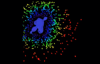
CASE_01
自然科学研究機構 生命創成探究センター
ImageXpress Micro Confocal
ハイコンテントイメージングシステム蛍光イメージングを用いた細胞内シグナル伝達系の研究
-
CASE_02
HCS Pharma
ImageXpress® Micro Confocal
ハイコンテントイメージングシステムハイコンテントイメージャー(HCA)とハイコンテントスクリーニング(HCS)アプローチを用いたマルチパラメトリック細胞イメージングアッセイ
-
CASE_03
Stemonix

SpectraMax i3x
マルチモードマイクロプレートリーダー試験管内でヒトの脳の発生と機能の均一性を高度に捕捉できるモデルが不足していた