モノクローナリティの検討とコンフルエンシーの評価のための、
様々な細胞に適用可能なラベルフリーと蛍光の自動イメージングソリューション
細胞株が単一の細胞に由来するモノクローナルであること、あるいは遺伝子が期待通りに編集されたことを証明することは、従来の技術では時間がかかり、非常に主観的なプロセスになりかねません。
CloneSelect™ Imagerは、高解像度の明視野イメージングにより、コンフルエンシーの測定およびモノクローナリティの検討を自動化できます。96ウェルプレートを2分未満でイメージングする能力は、業界トップクラスの画像取得速度です。単一細胞から形成されるコロニーのモニタリングは、バーコード付きのプレートを経時的に観察することで簡単に実施できます。また自動化された画像取得と解析により、正確で客観的、かつ一貫性のある結果が得られます。
CloneSelect Imager (CSI and CSI FL)の特長
-

迅速な単クローン性の確認
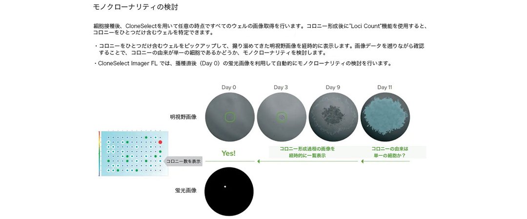
CloneSelect Imagerは96ウェルプレートをわずか2分以内でイメージングするという業界トップクラスの画像取得速度を誇ります。蛍光を用いて細胞播種0日目でモノクローナリティを確認することもできます。
-

マルチチャンネルイメージングとコンフルエンシーの自動判定
正確な細胞検出のために最適化されたアルゴリズムで、様々な細胞の種類や状態に対応します。そのまま公開することも可能な高解像度のイメージングにより、コンフルエンシーの自動解析とモノクローナリティの評価が可能です。
-

実証済みのスムーズなIND申請
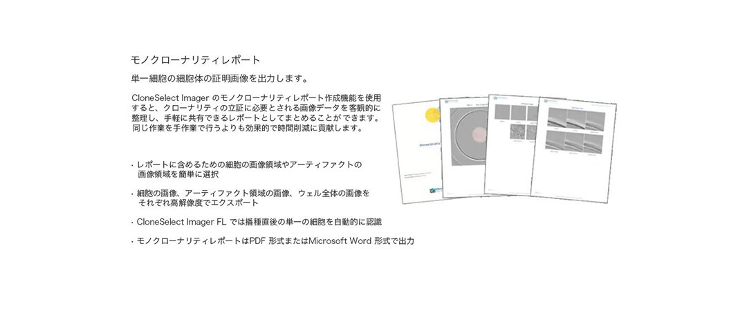
モノクローナリティレポートとは、FDAへの治験薬(IND)申請をサポートする監査対応文書を指します(21 CFR Part 312)。モノクローナリティレポート作成機能では、選択したパラメーターに基づいてレポートが自動的に生成されるため、FDAに提出する文書の作成が効率化されます。
-
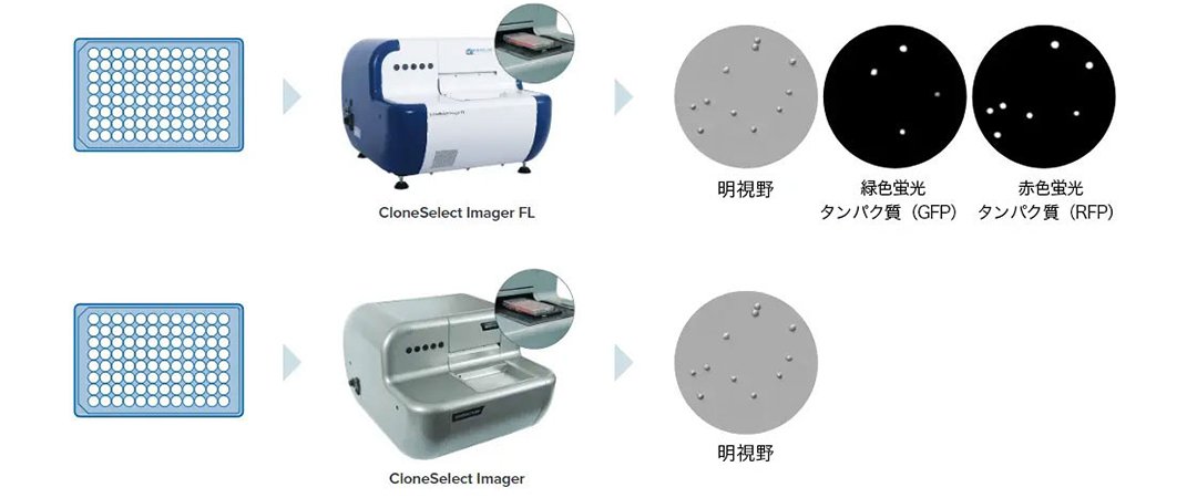
明視野および蛍光イメージング
標識不使用の明視野イメージングを用いて、迅速にすべてのプレートのすべてのウェルを画像取得します。マルチチャンネル蛍光イメージング(CSI FL)により、モノクローナリティアッセイの信頼性を高め、比較コンフルエンスアッセイ(赤色と緑色)を可能にします。
-
研究スケジュールを短縮するためのデータツール
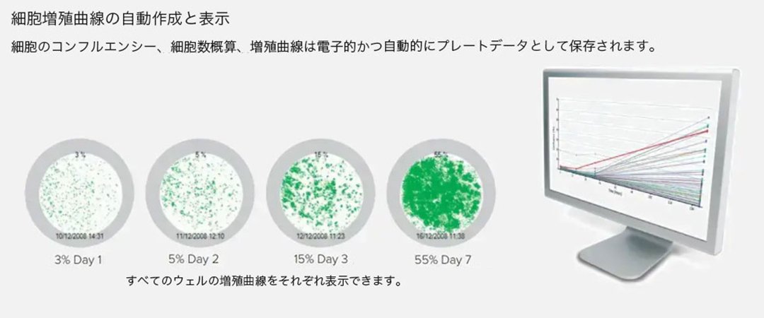
CloneSelect Imagerのソフトウェアは、細胞のコンフルエンシーを自動的に算出し、増殖曲線やヒートマップ、画像モンタージュを自動的に生成します。各ウェルの測定値は、自動的かつ経時的に追跡されます。これらの機能により治験薬申請文書提出のための細胞株樹立ワークフローの複数のステップ(イメージング、サンプル追跡、データ解析、レポート生成)を効率化します。
-
様々な細胞を迅速に画像取得
96ウェルプレートを2分以内に画像取得します。CHO 細胞、HEK 細胞、ハイブリドーマ、iPS 細胞といった多様な種類の接着性または沈降性懸濁細胞に対応します。
-
使いやすいソフトウェアによるインテリジェントな分析
ソフトウェアは各画像取得時点におけるコンフルエンシーを自動的に計算します。増殖曲線、画像モンタージュ、総増殖率、平均増殖率が自動算出され、データをエクスポートすることができます。ソフトウェアのガイドに従って操作するため、1時間程度の簡単なトレーニングで十分に使用することができるようになります。
-
高解像度イメージング
高速蛍光(CSI FL)および高解像度の明視野イメージングにより、細胞の残屑を含む単一細胞の正確な検出を可能にします。イメージングとデータのトラッキングは複数日にわたって行えます。
-
カスタマイズ可能な自動化オプション*
自動化・カスタマイズの専任チームが、ロボットによるプレートのローディングからインキュベーションなどを備えた完全自動化ワークステーションまで、様々なカスタムサービスを提供しています。
*価格、納期、仕様は相互に合意した技術的要件によって異なります。ソリューションの要件によって、標準性能に対し調整を行う場合があります。
合理化されたワークフロー(画像取得、解析、レポート)
(1) 画像取得
細胞をマイクロプレートに播種した後、培養中の任意のタイミングで画像取得します。
(2) 解析
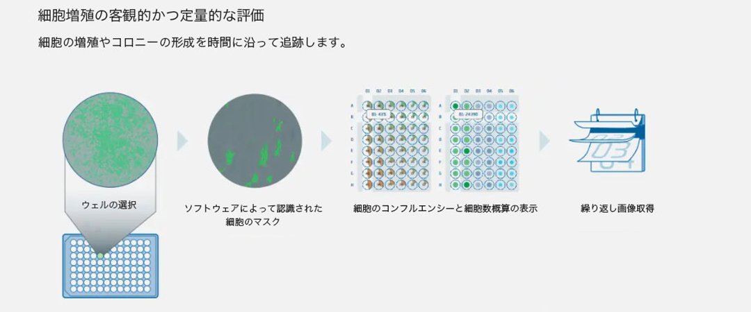
(3) レポート
詳細を見る
バーチャルデモ
詳細を見る
対応アプリケーション
-
細胞株開発
細胞株開発は、モノクローナル抗体などのバイオ医薬品分子を作製するプロセスにおいて重要なステップです。このプロセスは、多くの場合、目的の治療用タンパク質をコードするDNAを宿主細胞にトランスフェクションすることから始まります。高産生な希少細胞を単離するために何千ものクローンがスクリーニングされるが、これは手作業で時間のかかるプロセスです。
細胞株開発を見る > -
細胞生存率

細胞株開発には、標的治療タンパク質を高レベルで安定的に産生する単一細胞由来のクローンを発見することが必要です。このプロセスの重要な第一段階は、生存可能な単一細胞を単離することです。単一細胞は増殖してコロニーを形成し、標的治療タンパク質の生産性を評価することができます。単一細胞由来のクローンの生存率と増殖率は、CloneSelect ImagerとSpectraMax i3xマイクロプレートリーダーおよびイメージングサイトメーターで評価できます。
CloneSelect Imagerでのコンフルエンスと成長曲線の作成 >
EarlyTox Cell Integrity Kitによる細胞生存率と細胞毒性測定 >
CloneSelect Imagerでのコンフルエンスと成長曲線の作成 > -
コロニー形成アッセイ

軟寒天コロニー形成アッセイは、発がんの特徴であるアンカレッジ非依存性増殖を行う細胞の能力を特徴付けるための古典的で有名な手法です。半固形培地のようなコロニー形成を促進するマトリックスに播種した後、細胞は通常、コロニー増殖に影響を及ぼす可能性のある化合物とインキュベートされます。CloneSelect Imagerは、すべてのウェルを画像化し、コンフルエンスを自動的に計算し、コロニーの数と面積を推定し、コロニーの成長を追跡できます。
その他のアプリケーション
CloneSelect Imager (CSI and CSI FL)の仕様
| CloneSelect Imager | CloneSelect Imager FL | |
| イメージング | ||
| ソフトウェア | Windows 10 搭載の高性能なコンピューターにプリインストールされた専用の画像取得・解析ソフトウェア | Windows 10 搭載の高性能なコンピューターにプリインストールされた専用の画像取得・解析ソフトウェア |
|---|---|---|
| カメラ | 高解像度CMOSカメラ | 高解像度CMOSカメラ |
| 画像取得速度 | 90 秒(96 ウェルプレート) | 明視野:2 分以内(96/384 ウェルプレート) 蛍光:8 分以内(96/384 ウェルプレート) |
| 解像度 | 標準:3.6 μm | 高解像度:1.8 μm 標準:3.6 μm |
| 対物レンズ | 4x | 4x |
| 光源 | 明視野:白色透過光 光源:キセノンフラッシュランプ(5 W) 蛍光: 蛍光なし |
明視野:白色透過光 光源:キセノンフラッシュランプ(5 W) 蛍光:2 チャンネル(GFP、RFP) 光源:AURAIIIライトエンジン(LED) |
| 設置 | ||
| ソースプレートタイプ | 6、12、24、96および384ウェルのSBSマイクロプレートをラインナップ | 96ウェルおよび384ウェルのSBSマイクロプレート各種 |
| ソースプレート容量 | 1 xプレート | 1 xプレート |
| 設置重量 | 45kg(99ポンド) | 45kg(99ポンド) 外部光源は追加 3.6 kg(8ポンド) |
| ソースプレート容量 | 1 xプレート | 1 xプレート |
| 設置寸法 | 45.4cm(17.9インチ)×57.4cm(22.6インチ)×72.4cm(28.5インチ) | 45.4cm(17.9インチ)×57.4cm(22.6インチ)×72.4cm(28.5インチ) 外部光源:16.3 cm x 12.5 cm x 26.3 cm |
| 規制認可 | ||
| 適合規格 | CE | CE |
| 品質 | ISO9001:2008認証 | ISO9001:2008認証 |
詳細を見る
-
試薬・消耗品・アクセサリー
-
保守サービス・サポート