SpectraMax i3xマルチモードマイクロプレートリーダーの特長
-

蛍光性能の向上
キセノンフラッシュランプとLEDの強力な組み合わせにより、Spectral Fusion™照明で比類のない信号強度と優れた感度を実現しました。
-

ユーザーが柔軟にアップグレード、専用機並みの高性能を発揮
カートリッジを追加することでアルファスクリーン、時間分解蛍光、HTRF、インジェクターによる高速カイネティクス、ウェスタンブロット検出など、幅広い測定項目に対応しています。従来の光学系を改造して追加するシステムとは異なり、専用の光学系がカートリッジごとに内蔵されているため、各測定モード専用機並みの高性能を実現しています。
-

無染色細胞分析
明視野細胞セグメンテーション用のStainFree™ Cell Detection Algorithmにより、細胞に強いダメージを与える染色を行うことなく細胞計数とコンフルエント測定が可能になり、細胞計数ワークフローを簡素化することができます。
-
バックグラウンドノイズの低減と広いダイナミックレンジ
すべての励起波長にわたり感度を高めるためのSpectral Fusion™照射と、極めて微弱な光の検出を改善する冷却光電子増倍管(PMT)を搭載しました。希釈せずにより多くのデータを取得できます。
-
発光タイプのアッセイを簡単に捕捉
SmartInject™テクノロジーを搭載したSpectraMaxインジェクターカートリッジを追加することで、デュアルルシフェラーゼアッセイ、ATPアッセイといったフラッシュタイプの発光測定アプリケーションにも対応します。
-
マルチチャンネル機能
MiniMaxサイトメーターの2つの蛍光検出チャンネルは、ライブデッドやトランスフェクション効率などのレシオメトリックアッセイを含む、細胞生存率または細胞毒性アッセイの分析を可能にします。
-
細胞パスウェイ解析を1台で
細胞コンフルエンスと細胞生存率のイメージング、ウェスタンブロット解析、核酸とタンパク質の定量を1台のプレートリーダーで行うことができます。
-
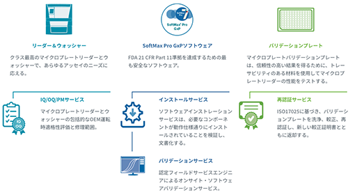
実績あるGxPソリューションでデータインテグリティとコンプライアンスを保証
GMP/GLP規制環境下施設のための当社の包括的な一連の実績あるコンプライアンスソリューションは、みなさまの取り組みを促進し、迅速に自信を持って、規制に準拠した施設を確立します。
- ・最高クラスのマイクロプレートリーダーおよび ウォッシャーですべてのアッセイニーズをサポート
IQ/OQ/PMサービスにより、規制を遵守したデジタルフォーマットで機器の文書を保存 - ・ソフトウェアインストールサービスで、操作仕様のために必要な要素がインストールされていることを確認し記録
- ・ソフトウェアバリデーションサービスはFDA 21 CFR Part 11ガイドラインをサポート
- ・IT担当者の負担を軽減するWindows Active Directoryとの連携やバリデーションプレートなどのトレーサブルな材料を用いて、信頼性のある結果を得るために必要なマイクロプレートリーダーの性能を試験
- ・最高クラスのマイクロプレートリーダーおよび ウォッシャーですべてのアッセイニーズをサポート
-
「SpectraDrop 微量サンプル測定プレート」でより少量のDNA、RNA、タンパク質をより迅速に定量
卓上型マイクロプレートリーダーのためのマイクロボリュームソリューション SpectraDrop™微量サンプル測定プレートは少量測定のためのハイスループットソリューションを実現します。革新的なデザインにより、サンプル調製時間を短縮し、2 µLという少量のDNA、RNA、タンパク質サンプルに対するラボの生産性を高めることができ、吸光、蛍光測定モードに対応します。マイクロプレートのフォーマットは均一で再現性のある分析を保証し、Molecular Devices 製StakMax®マイクロプレートスタッカーとシームレスに統合することで研究能力を向上させることができます。
詳しくはこちら
対応アプリケーション
-
吸光度

吸光度検出の仕組み、測定方法、濃度の算出方法など、吸光度検出のすべてをご紹介します。また、ELISA、核酸やタンパク質の定量、微生物の増殖など、一般的な吸光度アプリケーションやアッセイに関する情報も提供しています。
吸光度を見る > -
アルファスクリーン
AlphaScreen®テクノロジーは、分子内結合を検出するための均質でハイスループットな方法です。ドナービーズとアクセプタービーズが生体分子に結合し、ビーズが(結合によって)近接すると、化学反応のカスケードが起こり、SpectraMax® i3xとParadigm®マルチモードマイクロプレートリーダーで検出されるシグナルが増幅されます。
SpectraMaxパラダイムマルチモードマイクロプレート検出
プラットフォームでのAlphaLISAスクリーン > -
セルヘルス
細胞生存率とは、集団中の健康な細胞の数を指し、酵素活性、細胞膜の完全性、ATP産生、その他の指標を測定するアッセイを用いて評価することができます。これらの方法は、一般的な細胞生存能、あるいは特定の細胞経路の指標として、発光性、蛍光性、あるいは比色測定法を用いることができます。細胞毒性アッセイや細胞生存率アッセイは、薬剤や他の治療法の効果を評価するためにしばしば用いられ、新しい治療法を探索するための貴重なツールであると同時に、正常な細胞がどのように機能するかについての理解を深めるものでもあります。
セルヘルスを見る >
SpectraMax i3xマルチモードマイクロプレートリーダーの仕様
| 一般仕様 | |
| 読み取りモード | 吸光度 蛍光(上下読取式) 発光(上下読取式) 時間分解蛍光(上下読取式) 蛍光偏光(上下読取式) HTRF(上下読取式) アルファスクリーン(上下読取式) |
|---|---|
| 波長範囲 | Abs: 230 – 1000 nm FL Ex: 250 - 830 nm FL Em: 270 – 850 nm Lumi: 300 – 850 nm |
| 波長選択 | 1nm単位で調整可能なカートリッジフィルターとモノクロメーター |
| 吸光度測光精度/直線性 | < ±0.010 OD ±1.0%, 0 - 3 OD VIS 0 - 2 OD UV |
| 吸光度測光精度/再現性 | < ±0.003 OD ±1.0%, 0 - 3 OD VIS 0 - 2 OD UV |
| 上方測定の感度を最適化 | FL: Mono: 0.5 pM fluorescein 96 well, 1 pM fluorescein 384 well; Cartridge: 0.1 fmol/well in 75 μL fluorescein 384 well, 0.03 fmol/well in 8 μL fluorescein 1536 well Lumi: Mono: 3 pM ATP-Glow 96 well, 6 pM ATP-Glow 384 well; Cartridge: 20 amol ATP-flash TRF cartridge: 3 amol/well in 100 μL europium (0.03 pM) 384 well, 1 amol/well in 8 μL europium (0.125 pM) 1535 well FP cartridge: 3 mP (75 μl) fluorescein 384 well, 6 mP (8μl) fluorescein 1536 well AlphaScreen cartridge: <100 amol/well 384 well |
| 光源 | キセノンフラッシュランプ 超高出力LED |
| 検出器 | シリコンフォトダイオード 光電子増倍管 |
| 測定タイプ(エンドポイント、カイネティックなど) | エンドポイント カイネティック スペクトルスキャン ウェルスキャン |
| 読み取り時間 | Abs:29秒 FL:22秒 ルミ:21秒 TRF:21秒 FP:38秒 AS:22秒 |
| プレーティング | リニア 環状 |
| インジェクター | 〇 |
| イメージング | 〇 |
| ウェスタンブロット | 〇 |
| ロボティクス/オートメーション | 〇 |
| プレーティングタイプ | 6-1536 well plates |
| 温度制御 | 周囲温度+ 4°C to 45°C |
| 寸法 | H 32.5 cm x W 39.2 cm x D 60.55 cm |
| バリデーションツール | 吸光度バリデーションプレート 蛍光バリデーションプレート 発光バリデーションプレート マルチモードバリデーションプレート SoftMax® Pro GxPソフトウェア |
| 吸光度、蛍光およびルミネセンス | 〇 |
| 時間分解蛍光(TRF) | 〇 |
| 蛍光偏光(FP) | 〇 |
| Htrf | 〇 |
| アルファスクリーン | 〇 |
| SpectraMax® MiniMax® 300イメージングサイトメーターの技術仕様 | |
| 光源 | 特許取得済みソリッドステート照明、白色、460/20 nmおよび625/20 nm励起 |
| 検出器 | 1.25メガピクセル、12ビット高感度CCDカメラ |
| 発光 | 明視野; 緑 541/108 nm; 赤 713/123 nm |
| 対物レンズ | シングル4倍対物レンズ |
| オートフォーカス | 特許取得済みレーザースキャニングオートフォーカス |
| 解像度 | 1.9 μm x 1.9 μm ピクセルサイズ |
| データ収集・解析ソフトウェア | SoftMax® Proソフトウェア MiniMax Imaging Edition |
| 画像取得速度* (1色 - 96ウェル) | 取得 3:40 取得 + 解析 6:30 |
| 試料キャリア | ANSI/SBS準拠マイクロプレート 96および384ウェル |
| 寸法(cm) | 39.2 cm 幅 x 19.5 cm 高さ x 60.6 cm 奥行き(SpectraMax® MiniMax™ 300イメージングサイトメーター) 39.2 幅 x 44.0 高さ x 60.6 長さ (ベースシステム付き) |
詳細を見る
-
試薬・消耗品・アクセサリー
- EarlyTox心毒性キット
- EarlyTox細胞生存率アッセイキット
- SpectraMax Quant dsDNAアッセイキット
- CatchPointシンプルステップELISAキット
- CatchPoint cAMP蛍光アッセイキット
- CatchPoint cGMP蛍光アッセイキット
- FLIPRカルシウムアッセイキット
- FLIPR カリウムアッセイキット
- SpectraMax DuoLucレポーターアッセイキット
- SpectraMax Glo Steady-Lucレポーターアッセイキット
- 神経伝達物質トランスポーター取り込みアッセイキット
- QBT脂肪酸取り込みアッセイキット
- ScanLater ウェスタンブロットアッセイキット
-
保守サービス・サポート
導入事例
-
CASE_01
佐賀大学 農学部生命機能科学科

SpectraMax i3x
マルチモードマイクロプレートリーダー多検体ハイスループットスクリーニングによる酸化ストレス応答転写因子の研究
-
CASE_02
iTeos Therapeutics SA
SpectraMax i3x
マルチモードマイクロプレートリーダーi3xはラボで実施されるさまざまなアッセイに絶妙にアダプターできる点が気に入っています
-
CASE_03
Ionic Transport Assays Inc.
SpectraMax i3x
マルチモードマイクロプレートリーダー幹細胞由来心筋細胞を用いた薬剤候補により誘導される心毒性の評価