大型のオルガノイドやスフェロイドの3次元(3D)画像を
最大2倍の速さで画像取得
ImageXpress® Confocal HT.aiハイコンテントイメージングシステムは、8つのイメージングチャンネルに対応する7波長のレーザー光源を利用して、短い露光時間でハイスループットな画像取得を可能にすると同時に、高度なマルチプレックスアッセイを実現します。水浸対物レンズが画像の解像度を高め、光学収差を最小限に抑えることで、厚みのあるサンプルをより深く観察できるようになります。
MetaXpress®ソフトウェアとIN Carta™ソフトウェアを効果的に組み合わせれば、機械学習機能と直感的なユーザーインターフェースにより、高度な表現型の分類と3D画像解析のワークフローを簡略化できます。
ImageXpress Confocal HT.ai
ハイコンテントイメージングシステムの特長
-

アッセイのフレキシビリティをさらに高度化
レーザー励起による8つのイメージングチャンネルにより、アッセイのフレキシビリティ、画像の輝度、QuickIDといったターゲット画像取得のフレキシビリティが向上しました。自動水浸対物レンズは開口数が大きく、サンプルと浸液の屈折率が一致しており、高解像度と低収差を両立しています。
-

ハイスループットで高品質な画像
高出力な励起光源によって、シグナル輝度の増加、露光時間の短縮、3Dサンプルの高速な画像取得を実現しました。マイクロレンズで強化されたスピニングディスク型共焦点光学系は視野がフラットで、より正確で再現性の高い画像解析を実現します。露光時間が短縮され、スキャン速度が最大で2倍高速化するほか、CFPおよびYFP用レーザーを用いたFRETの実験も実施でき、研究の幅を広げます。
-

IN Carta画像解析ソフトウェア
機械学習を活用して、ハイコンテントイメージングの解析精度と頑健性を向上し、他の技術では得ることができないデータの見識を提供します。Phenoglyphsモジュールは頑健で学習可能な分類機能を提供し、SINAPモジュールはあらゆる種類の画像に対して学習可能なセグメンテーション機能をもたらします。
-
高輝度レーザー光源
高出力レーザーによる励起で露光時間を最大75%減少させることができます。近赤外波長を含む7波長のレーザー光源は、多重染色アッセイを行うユーザーに最適です。
-
唯一無二のAgileOptix™スピニングディスクテクノロジー
独自の光学系、高出力レーザー光源およびsCMOSセンサーにより、優れた感度を実現します。交換可能なディスクモジュールにより、高速な画像取得と高解像度な画像取得を柔軟に切り替えることができます。
-
多数のイメージングモード
標準で蛍光、ワイドフィールド、および共焦点イメージングを搭載しています。オプションでは位相差および明視野ラベルフリーイメージング、水浸光学系に対応できます。
AgileOptix™テクノロジー
AgileOptixテクノロジーは、パワフルな固体光源、カスタム光学系、sCMOSセンサー、異なるディスクモジュールを交換できる機能を組み合わせています。
同じ露光時間で画像取得した場合の輝度の比較
IN Carta画像解析ソフトウェア
直感的なユーザーインターフェースとパワフルな解析機能を組み合わせることで、高度な表現型の分類と3D画像解析のためのワークフローがシンプルになります。高度な機能により、データを大規模に解析し、複雑な前処理や後処理を必要とせずにリアルタイムの見識を提供します。オプションのPhenoglyphsモジュールに搭載された機械学習は、複雑な分類を自動的に実行します。
詳しくはこちら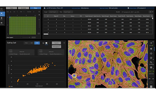
詳細を見る
フレキシブルなイメージングソリューションを使って
研究を拡張できます
モレキュラーデバイスは、ImageXpress Confocal HT.aiシステムに対して、フレキシブルなオプションを提供しています。丸底あるいは平底プレートといった様々なサンプルフォーマットでの画像取得や、環境制御下での細胞のモニタリングなど、様々な研究ニーズを満たす柔軟性の高いシステムです。30年を超えるイメージングの経験を持つ当社が、アッセイに最適な画像を確実かつ正確に取得することのできるオプションの選択をお手伝いします。
標準のハードウェアオプション
 水浸対物レンズ
20倍、40倍、60倍の水浸対物レンズを用いると、光学的収差が改善され、深さ方向の画像精度が向上し、短い露光時間でも明るい画像を取得できます。
 環境コントロールユニット
環境コントロールユニットを用いると、培地の蒸発を最小限に抑えながら温度と湿度を維持でき、複数日に渡る生細胞のタイムラプスイメージングが行えます。
 透過光ユニット
当社の透過光ユニットと位相差対物レンズを用いると、バックグラウンドと容易に区別できる、コントラストの高い非染色細胞の位相差観察像を簡単に取得できます。
特注・カスタマイズのオプション
モレキュラーデバイスは、ImageXpress Confocal
HT.aiシステムをカスタマイズして、ユーザーのニーズに合わせた調整を行うことができます。インキュベーター、リキッドハンドラー、フルオートメーションワークセル用のロボットなどの他社の装置とも統合できます。ライフサイエンス業界で30年以上の経験を持つ当社は、高品質な製品と世界中でのサポートを提供しています。
販売は当社のCustom Product Purchase Terms(www.moleculardevices.com/custom-products-purchase-terms(USサイトへ遷移します))に従って行います。
 深部組織観察用共焦点ディスクモジュール
深部組織観察用共焦点ディスクモジュールは、クロストークを低減させて焦点外の光ノイズを抑制し、組織のより深い部分に到達するクリアな観察を可能にします。
ハイスループットな長期カイネティクス
温度、O₂(低酸素)、CO₂、および湿度を一定条件に保ちながら、複数枚のプレートを長期間にわたって画像取得するスケジュールを組むことができます。生細胞観察できるプレート枚数を200プレート以上に拡張できます。
 ロボティックオートメーション
スループットを高め、ヒューマンエラーをなくし、無菌状態を維持しながら、一貫したサンプル処理を行います。モジュールで構成要素を追加できる設計であり、アップグレード可能です。

深部組織観察用共焦点ディスクモジュール
レーザー光源と組み合わせて使用する特別な深部組織観察用共焦点ディスクモジュールは、厚みのある組織サンプルを画像取得する際の分解能を改善できます†。 †データと画像は顧客サンプルを使用して開発中に得られたものです。結果は異なる場合があります。製品の価格、納期、および仕様は、双方が合意した技術要件に応じて変更される場合があります。ソリューション要件に応じて標準性能が調整される場合があります。
- ・焦点外の光ノイズを低減
- ・ヘイズ(ピンホールクロストーク)を低減
- ・厚みのある組織サンプルの深くまで透過し、シャープな画像を取得
詳細を見る
画像ギャラリー


†データと画像は顧客サンプルを使用して開発中に得られたものです。 結果は異なる場合があります。 製品の価格、納期、および仕様は、双方が合意した技術要件に応じて変更される場合があります。 ソリューション要件に応じて標準性能が調整される場合があります。
対応アプリケーション
-
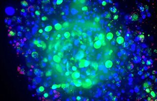
3D細胞イメージングと解析
3次元(3D)細胞モデルは、現在非常に注目されており、in vivoで生じている組織の微小環境や細胞間相互作用、生物学的プロセスをより生理学的に正しく再現できるとされています。ImageXpressシステムのようなハードウェアテクノロジーとMetaXpress®ソフトウェアの統合された3D解析モジュールを組み合わせることで、予測能の高いデータを生成できます。このインターフェースだけで、スループットやデータの質を犠牲にすることなく、3D画像取得と解析の課題に応えることができ、発見に信頼性をもたらします。
3D細胞モデルを見る > -
セルペイント
セルペイントは、ハイコンテントな多重染色画像を利用したアッセイで、細胞プロファイリングに利用されます。セルペイントアッセイでは、最大6種類の蛍光色素が使用され、核、小胞体、ミトコンドリア、細胞骨格、ゴルジ体、RNAなどの様々な細胞の成分を標識します。これは、細胞全体の代表的な画像を取得するために、できるだけ多くの細胞を「ペイントする」ために行われます。
セルペイントを見る > -
疾患モデル
疾患モデル系は、単純な2次元細胞培養から複雑なモデル生物まで、その複雑さと規模は多岐にわたります。モデル生物はin vivoの状況を提供する一方で、コストがかかることが多く、ヒトの生物学を表現できないことがあります。一方、従来の2次元細胞培養システムは長年使用されてきたが、生体組織に見られる複雑な3次元構造や細胞間相互作用を表現するには限界がありました。その結果、3次元細胞培養は、疾患モデル化のための魅力的なモデル系として浮上してきました。
疾患モデルを見る >
ImageXpress Confocal HT.ai ハイコンテントイメージングシステムの仕様
| カメラ | sCMOS(4メガピクセル以上) |
|---|---|
| 視野面積 | 1.96 mm²(対物レンズ10倍使用時) |
| サンプル処理速度 | 200,000ウェル/日以上 |
| 対物レンズ | ドライ(1X~100X)、油浸(40X、60X、100X)および水浸(20X、40X、60X) |
| フィルター切替方式 | 8ポジションの吸収フィルターホイールおよび5ポジションのダイクロイックミラーホイール |
| オートフォーカス方式 | レーザーオートフォーカス、イメージオートフォーカス(併用可能) |
| 位相差観察(光学式) | ◯ |
| 透過光観察 | ◯ |
| 明視野観察 | ◯ |
| サポートされるプレート | 1536ウェルプレートまで |
| スライドガラスの観察 | ◯ |
| ロボティクス・自動化への適合性 | ◯ |
| 温度制御オプション | ◯ |
| CO₂制御オプション | ◯ |
| リキッドハンドリングおよび環境コントロールオプション | ◯ |
| 共焦点観察 | ◯ |
| ワイドフィールド | ◯ |
| ソフトウェア | MetaXpress画像取得・解析ソフトウェア、IN Carta画像解析ソフトウェア |
| MDCStoreデータマネージメント | ◯ |
| カスタムモジュールエディター | ◯ |
詳細を見る
-
試薬・消耗品・アクセサリー
該当の試薬・消耗品はありません。
-
保守サービス・サポート