FLIPRカルシウム測定キット
FLIPRカルシウム測定キットとは
FLIPR®カルシウム測定キットは、創薬や基礎研究のための細胞内カルシウムの変化を測定するための最適なプラットフォームです。
・細胞洗浄アーチファクトを低減することで、幅広い種類の細胞からの蛍光シグナルを最大化します。
・ウェル間のばらつきを低減し、ハイスループットスクリーニングのアッセイ品質(Z'ファクター)と信頼性(CV %)を向上
FLIPRカルシウム測定キット
- ・普遍的な添加と測定のワークフローにより、アッセイのワークフローを加速し、スループットを向上させます。
- ・優れたシグナル対ノイズ比により、アッセイ開発中の内因性または一過性にトランスフェクトされたレセプター活性の確認が容易
- ・FLIPR® TetraおよびFlexStation® システム用に最適化・検証済みのプロトコールにより、ルーチンから非定型の細胞株やターゲットまで対応可能
カルシウム試薬の最も包括的なポートフォリオとして構築されたこのキットは、最適化済みの均一性蛍光ベースの製剤を提供し、GPCRおよびイオンチャネルのターゲットのアッセイ開発とスクリーニングを迅速化します。FLIPRカルシウム測定キットは、シンプルで信頼性の高い均一性アッセイフォーマットで、細胞内カルシウム変化を検出するための最も包括的な方法を提供します。6種類の製剤から目的のターゲットを補うものをお選びください。
FLIPRアッセイキットは、蛍光のバックグラウンドノイズを低減し、シグナル対ノイズ比を改善するために消光色素を採用しています。特許取得済みのクエンチ技術は、FLIPRアッセイキットの購入を通じて、創薬研究者およびライフサイエンス研究者にモレキュラーデバイスが独占的に提供しています。
主な特徴と利点
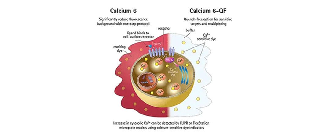
FLIPR Calcium 6 Assay Kit、FLIPR Calcium 6-QF Assay Kit
カルシウムアッセイキットの中で最大のシグナルウインドウを提供します。シグナル振幅の大幅な増大は、内因性、初代、幹細胞ターゲットなどの低シグナルスクリーニングの再開に役立ちます。さらに、この新規色素は有機陰イオン交換タンパク質の影響を受けないため、プロベネシドをほとんど含まない状態での測定が可能である。
クエンチフリー(QF)製剤で使用する場合、この色素はクエンチに敏感なターゲットや、1ウェル内でカルシウム流動アッセイと発光アッセイのマルチプレックス要件が必要なアッセイに最適です。
FLIPR Calcium 5 Assay Kit
FLIPR Calcium 4 Assay Kitで使用されている優れたマスキング技術を活用し、バックグラウンド蛍光を低減しながら、アッセイシグナルウィンドウを増幅する新規蛍光色素です。上記のアッセイに加え、レセプターの発現が低いレセプターや小さなカルシウム応答を示すその他のターゲットも本キットの候補となります。
FLIPR Calcium 4 Assay Kit
細胞外溶液中の消光性を高めた改良型特許取得済みマスキング色素を配合し、バックグラウ ンドを大幅に低減し、シグナルウィンドウを拡大。より不活性で、化合物の干渉を低減します。カルシウムチャネル、ケモカインレセプター、トランスフェクタントレセプターなどがターゲットです。
FLIPRカルシウム測定キットのデータ
-
FLIPR Calcium 6 Assay Kitシグナルウィンドウ
CHO M1細胞におけるムスカリンM1受容体のカルバコールアゴニズムにおけるカルシウムフラックスに対する蛍光シグナル応答の比較。FLIPR Calcium 6 Assay Kitを用いた培地中均一性アッセイによるシグナル対ノイズ比を示す。FLIPR Calcium 6 Assay Kitは、新規の蛍光色素と実績のあるクエンチ技術を組み合わせることで、最高のSB比を示します。 -

クエンチャーフリーのシグナルウィンドウ
CHO M1細胞におけるムスカリンM1受容体のカルバコールアゴニズムにおけるカルシウムフラックスに対する蛍光シグナル応答の比較。FLIPR Calcium 6 Assay Kitを用いた培地中均一性アッセイによるシグナル対ノイズ比を示す。FLIPR Calcium 6 Assay Kitは、新規の蛍光色素と実績のあるクエンチ技術を組み合わせることで、最高のSB比を示します。 -
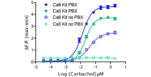
プロベネシドフリーシグナルウィンドウ
細胞株によっては有機陰イオン交換タンパクに対する耐性が高い。プロベネシドを含まないFLIPR Calcium 5 Assay Kit(Ca5キット、PBXなし)を使用した場合、シグナルは認められません。FLIPR Calcium 6 Assay Kit(Ca6)では、プロベネシドを使用しないためシグナルは小さくなりますが、EC50は同等であり、Z'は0.5以上です。 -
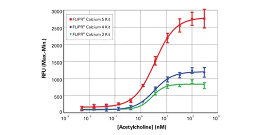
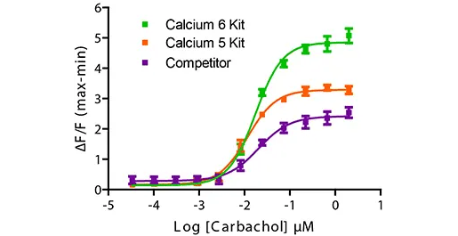
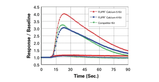
FLIPR Calcium 5、4、3 Assay Kitの比較
CHO M1細胞におけるムスカリンM1受容体のアセチルコリン作動時のカルシウムフラックスに対する蛍光シグナル応答の比較。クエンチ技術を用いたFLIPRカルシウム測定キットのシグナル対ノイズ比を示します。FLIPR Calcium 4 Assay Kitの新規蛍光色素と実績のあるクエンチ技術を組み合わせたFLIPR Calcium 5 Assay Kitは、従来のキットよりも高いSB比を示します。 -
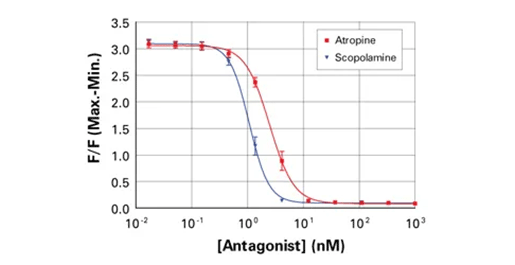
FLIPR Calcium 5 Assay Kitによる拮抗作用データ
FLIPRカルシウム5アッセイキットとFLIPRテトラシステムを用いて、CHO M1細胞のEC80アセチルコリンに対するカルシウム流動アッセイの拮抗作用を評価。 -
シグナルトレースの比較
異なるカルシウム動員キットを用いて、HEK 293細胞におけるPY受容体のATPアゴニズムをFLIPR Tetraシステムで比較したシグナルトレース。すべてのキットで一貫したEC50値が記録され、バックグラウンドの増加は見られなかった。FLIPR Calcium 5 Assay Kitでは、シグナルウィンドウが大幅に拡大した。 -
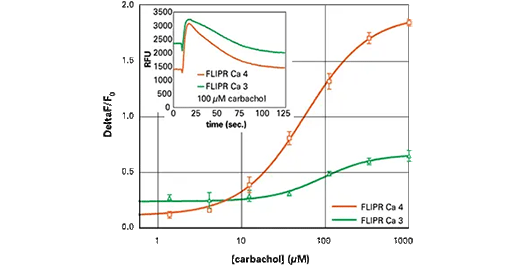
FLIPR Calcium 4 Assay Kitと3 Assay Kitの比較
FLIPR Calcium 4 Assay Kit(オレンジ)は、FLIPR Calcium 3 Assay Kit(緑)に比べ、バックグラウンドが減少し、ΔF/Fo値が改善しました。アッセイ 384ウェルプレート、10,000細胞/ウェル、FLIPR Tetraシステムでのアッセイ前に培地を除去したHEK 293細胞におけるカルバコールに対する内因性応答。 -
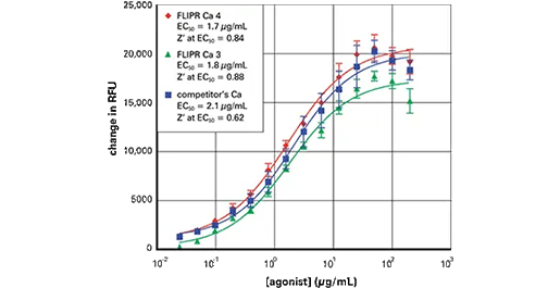
FLIPR Calcium 4および3 Assay Kitと競合キットの比較
多くの場合、FLIPR Calcium 4 Assay KitとCalcium 3 Assay Kitは同等の性能を示し、同時に競合キットを凌駕します。アッセイ アッセイ:タンパク質活性化剤で刺激した非固着細胞、培地除去なし、アッセイはFLIPR 3インストゥルメンテーションで実施。
FLIPRカルシウム測定キットの技術
-
FLIPRカルシウム測定キットはGPCRとイオンチャネルの評価にホモジニアスなソリューションを提供します。
FLIPRカルシウム測定キットは、インキュベーション中に細胞質に吸収されるカルシウム感受性色素を利用します。リガンドとレセプターが結合すると、細胞内のカルシウムが放出され、色素と結合して蛍光強度が増加します。すべてのFLIPRカルシウム測定キットは細胞外マスキング技術を利用してバックグラウンド蛍光を遮断し、アッセイのシグナルウィンドウを拡大します。FLIPR Calcium 6-QF Assay Kitは例外的に、マスキング技術を使用せずにアッセイを行うことができるため、発光アッセイとのマルチプレックスなど、クエンチに敏感なアプリケーションに最適です。
FLIPRカルシウム測定キットに対応する製品・サービス
-
SpectraMax iD3/iD5 マルチモードマイクロプレートリーダー
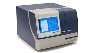
大型タッチスクリーンを備えた高感度マイクロプレートリーダー
-
FlexStation 3 マルチモードマイクロプレートリーダー
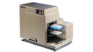
イオンチャネル・GPCR活性の測定に威力を発揮する
自動マルチチャンネルピペッター搭載マルチマイクロプレートリーダー -
SpectraMax i3x マルチモードマイクロプレートリーダー
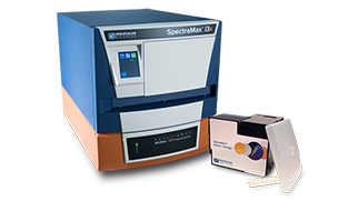
研究ニーズに合わせて進化できるマイクロプレートリーダー
-
FLIPR Penta ハイスループットセルベーススクリーニングシステム
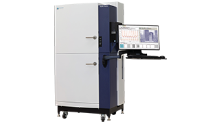
リード化合物の同定および化合物の安全性評価を目的とした
ハイスループットカイネティックスクリーニングに理想的なシステム