EarlyTox Cell Integrity Kit
EarlyTox Cell Integrity Kitとは
EarlyTox™ Cell Integrity Kitは、蛍光標識により生細胞と死細胞の識別を可能にします。これは、SpectraMax® MiniMax™ 300イメージングサイトメーターやその他の蛍光ベースの細胞イメージング装置と併用することで、細胞生存率の迅速な定量化に役立ちます。
EarlyTox Cell Integrity Kit
EarlyTox™ Cell Integrity Kitは、生細胞と死細胞の同定を簡便化する最適化された試薬セットです。このキットは、細胞生存率に対する様々な処理の効果を測定し、アポトーシスやネクローシスを含む様々なメカニズムを介した毒性効果を評価するために使用できます。このキットは、付着性、非付着性を問わず、多くの細胞種で使用できるように設計されている。プロトコールはシンプルなワークフローと試薬の性能により、ハイスループットスクリーニングに適している。
・高いシグナルにより、短時間で結果が得られる
・多くの細胞種に対応するよう設計
・単一のシステムで画像取得と解析を合理化
EarlyTox Cell Integrity Kitのデータ
-
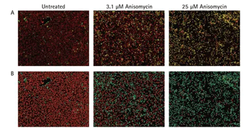
未処理および化合物処理したHeLa細胞の画像
(A)未処理細胞(左パネル)は、ほとんどが生きており、核は赤色蛍光を発している。中間濃度の化合物(中央パネル)では、生細胞と死細胞が混在している。高濃度の化合物(右パネル)では、ほとんどの細胞は死んでおり、核は赤と緑の両方で標識されている。画像はSpectraMax® MiniMax™ 300イメージングサイトメーターで取得した。(B) SoftMax® ProソフトウェアのClassification機能を用いて、細胞を生細胞(赤色マスク)または死細胞(青色マスク)に識別した。 -
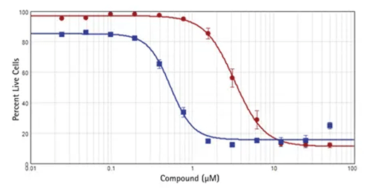
細胞毒性化合物のIC50曲線
(A)HeLa細胞をアニソマイシン(赤丸)またはスタウロスポリン(青四角)で処理した。結果は、SoftMax® Proソフトウェアの4パラメータ曲線フィットを用いてプロットした。アニソマイシンのIC50は3.3μMで、スタウロスポリンのIC50は0.53μMであった;両値とも公表値と一致していた。
EarlyTox Cell Integrity Kitの技術
-
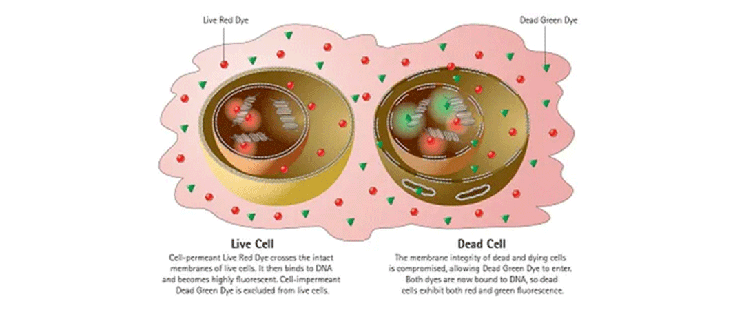
アッセイキットのメカニズム
EarlyTox Cell Integrity Kitは2種類の核色素を使用し、細胞の損傷や壊死、アポトーシス、その他のメカニズムによる細胞死による細胞外膜透過性の変化を検出することができます。細胞生存率は、生細胞と損傷細胞の数またはパーセンテージをカウントすることにより評価される。このアッセイは、細胞生存率に対するさまざまな処理の影響の研究、医薬品化合物やその他の化学物質の毒性効果の評価、ネクローシスやアポトーシスの研究、その他多くの用途に使用できる。