Mimetas, The Netherlands
企業/大学
Mimetas
使用製品
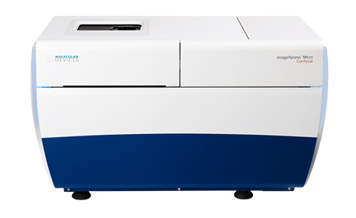
ImageXpress Micro Confocalハイコンテントイメージングシステム
ImageXpress Pico自動細胞イメージングシステム
ImageXpress Micro 4ハイコンテント イメージングシステム
臓器チップ企業
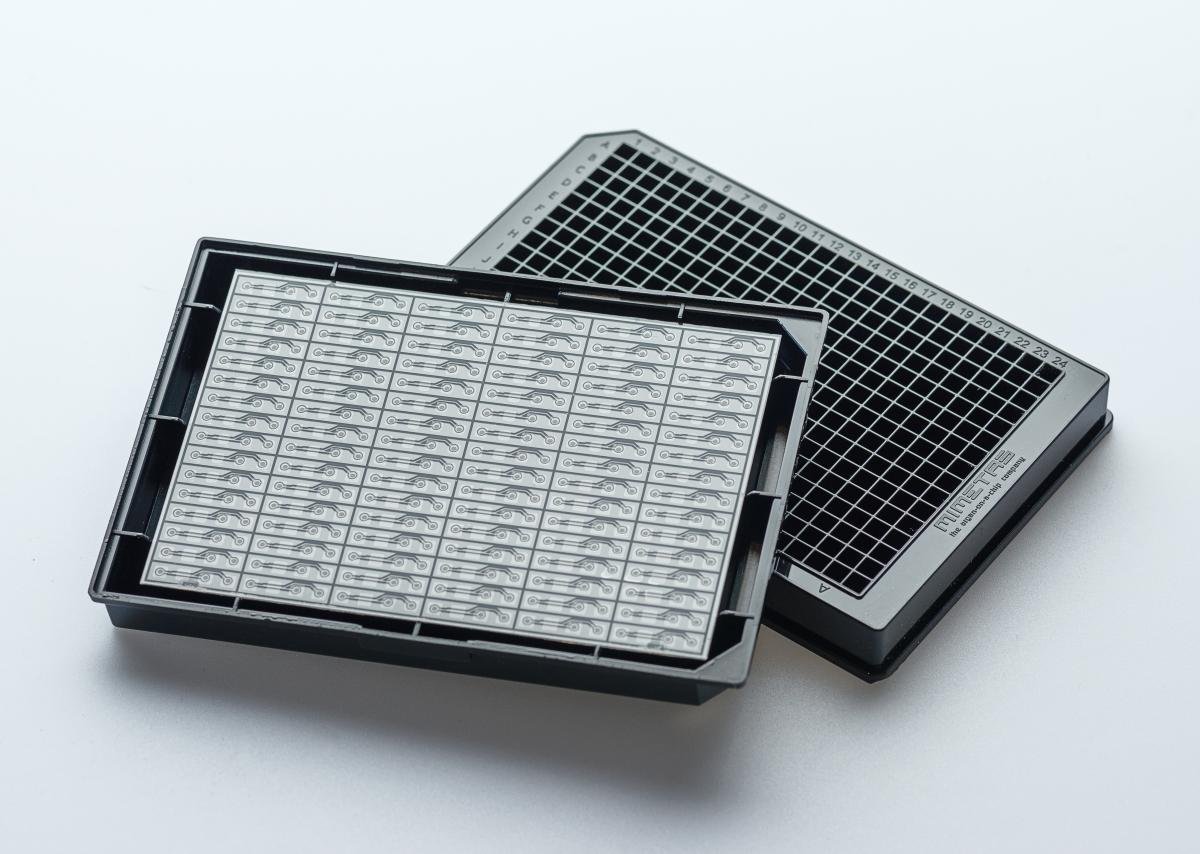
MIMETASはユニークな3D臓器on-a-chipプラットフォームであるOrganoPlate®を提供している。OrganoPlate®は完全に互換性のあるマイクロ流体培養プレートであり、小型化された臓器モデル上であらゆるスループットでの化合物のテストを可能にする。OrganoPlate®は、連続灌流下での3Dセル培養、メンブレンフリーの共培養、バウンダリーやグラジエント形成をサポートし、組織や臓器の重要な側面を模倣することで、より優れた組織モデルや疾患モデルの予測につながります。OrganoPlate®をベースに、MIMETASは製薬業界向けにカスタム組織モデルを開発し、新薬や個別化治療のスクリーニングと開発を迅速化・改善する。OrganoPlate®のモデルの概要は、Molecular Devices社主催の最近のウェビナーで見ることができる。
MIMETASでは、OrganoPlate®を研究、開発、薬剤スクリーニングのための疾患、毒性、輸送モデルの開発に活用している。脳モデル、腎臓毒性モデル、癌モデル、肝臓モデル、腸モデル、内皮血管系モデルなど、様々な組織モデルが開発されている。これらの血管系モデルは、他の組織培養と組み合わせて、血管系組織モデルを確立することができる。
組織モデルの開発と応用には、位相差タイムラプス、蛍光顕微鏡、共焦点顕微鏡など、さまざまな画像ベースの読み出しと、自動化された3D画像解析を組み合わせて使用する。チームはImageXpress Microコンフォーカルハイコンテントイメージングシステムを日常的に使用しており、ほとんどのインチップアッセイで標準的なハイスループット画像装置として活躍している。
ミメタスのマネージング・ディレクター兼共同設立者であるJos Joore氏は、「ImageXpress Micro Confocalは、我々の3D Organ-on-a-chip プラットフォームを真のハイスループット・システムとして使用することを可能にします。ImageXpress MicroとImageXpress Micro ConfocalはどちらもOrganoPlate®に完璧にマッチします。"
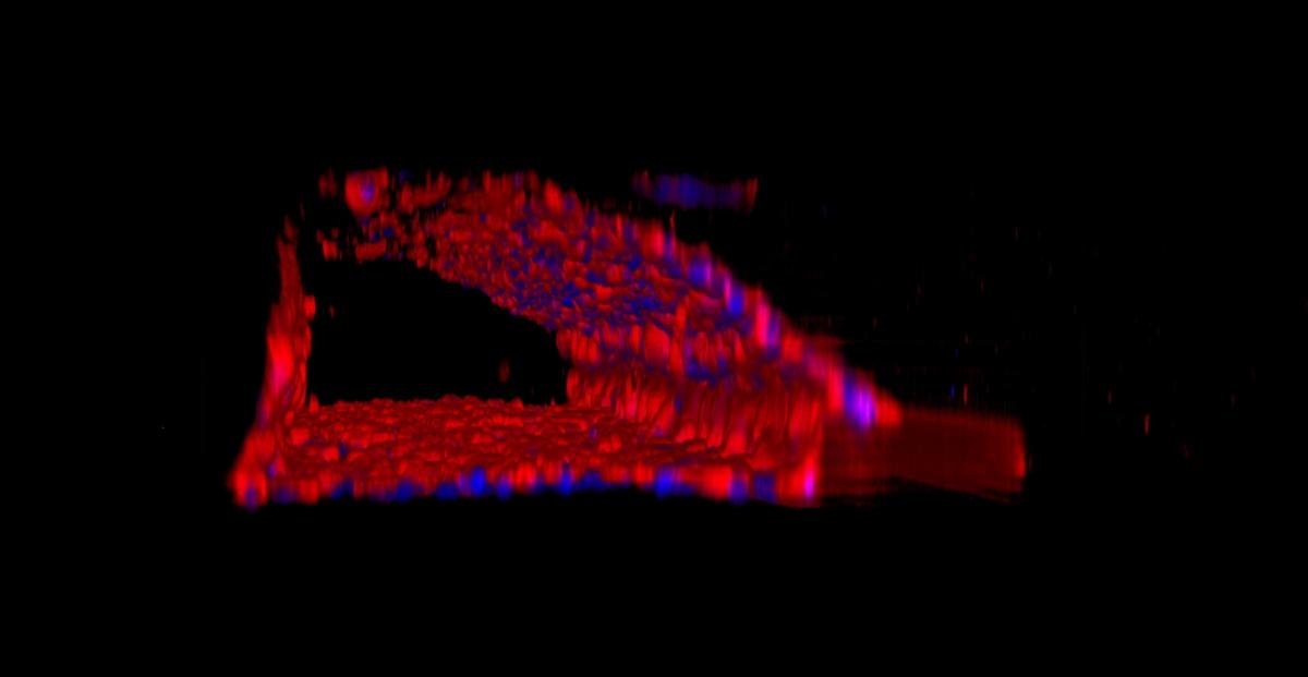
MIMETAS OrganoPlate®におけるマイクロ流体腎臓近位尿細管モデル。
ヒト腎臓細胞(Sigma RPTEC)をコラーゲン1に播種し、Ezrin(赤)とDapi(青)を染色した。Ezrinは近位尿細管の先端側の微絨毛を表す。
参考文献
Wevers, N.R. et al. High-throughput compound evaluation on 3D networks of neurons and glia in a microfluidic platform. Nature Scientific Reports 6, Article number: 38856 (2016).
Junaida A. et al. An End-User Perspective on Organ-on-a-Chip: Assays and Usability Aspects. Current Opinion in Biomedical Engineering. Article in Press (2017).