- Overview
- Data
- Technology
- Resources
- Specification
Key advantages:
- The semi-solid CloneMedia method of cloning prevents the overgrowth of fast-growing clones, allowing for the selection of potentially valuable slow-growing clones
- Hands-on time can be reduced when the workflow is combined with a ClonePix system. Hypoxanthine-aminopterin-thymidine (HAT) selection and cloning of hybridomas are accomplished in one step, minimizing the required time and materials
- Ready to use (no extra addition of supplements required)
Our XP Media and CloneMedia Complete Kit for Mouse Hybridoma Generation (P/N K8861) is composed of the following components. All components can be sold separately.
| COMPONENT | DESCRIPTION | |
|---|---|---|
| XP Media Pre-Fusion Myeloma Growth Medium and Hybridoma Expansion Medium (without HT) P/N K8862 | 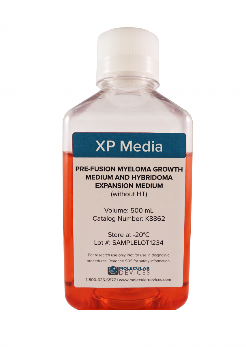 |
Supports the growth of myeloma cells before fusion. Also supports expansion of hybridoma clones. Does not contain hypoxanthine or thymidine (HT). |
| XP Media Hybridoma Fusion Medium P/N K8863 |  |
Used to wash cells before fusion and during fusion process. Does not contain supplements to support growth. |
| XP Media Hybridoma Fusion Recovery Medium P/N K8864 | 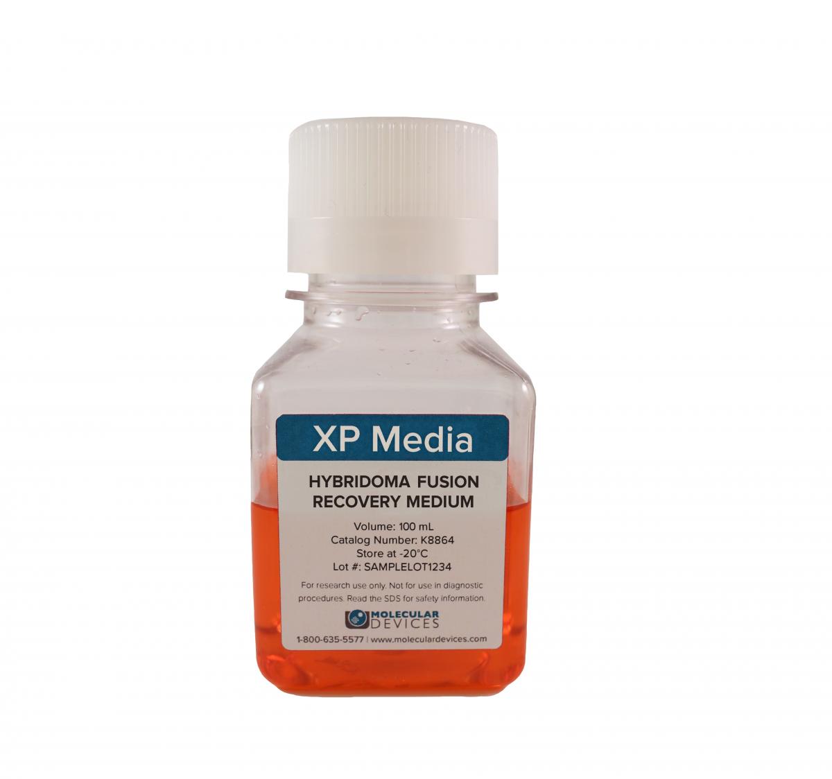 |
Used to promote hybridoma viability after the fusion process but before clone selection. Does not contain hypoxanthine, aminopterin, and thymidine (HAT). |
| CloneMedia Hybridoma Semi-Solid Selection and Cloning Medium (with HAT) P/N K8865 |  |
Used after fusion of splenocytes and myeloma cells to select and clone hybridomas in one step. Methycellulose-based formula optimized for colony formation. Equally suitable for fresh fusions and for stable hybridoma cell lines. |
| XP Media Hybridoma Growth Medium (with HT) P/N K8866 | 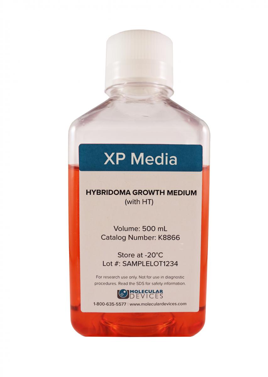 |
Optimized for hybridoma expansion following clone selection and colony picking. Contains hypoxanthine and thymidine (HT) and is used to wean hybridomas off aminopterin from the selection process. |
| Hybridoma Polyethylene Glycol (PEG) for Cell Fusion P/N K8868 | 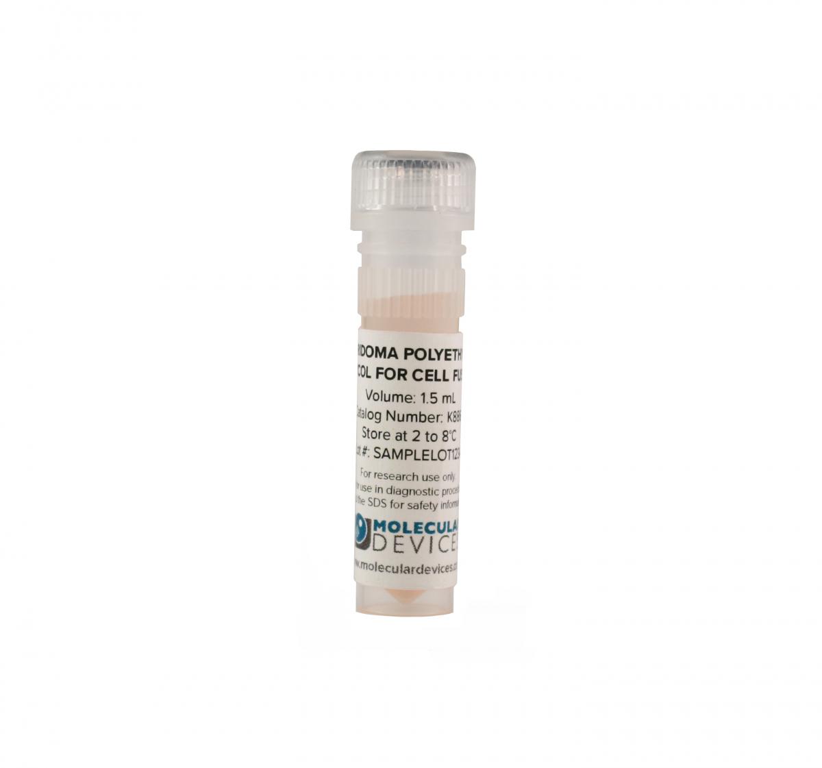 |
Used for the fusion of mouse splenocytes and parental myeloma cells to generate hybridomas. PEG is present as a 50% solution in DMEM. |
| ALTERNATIVE HYBRIDOMA SEMI-SOLID SELECTION AND CLONING MEDIUM | ||
|---|---|---|
| CloneMedia Hybridoma Semi-Solid Selection and Cloning Medium (without HAT), P/N K8867 | 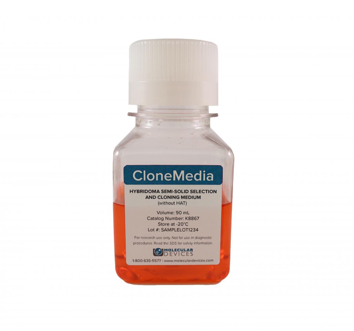 |
If you are using alternative hybridoma selection methods, then this medium will be perfect for your needs. Researchers can add desired selection reagents into this medium and use to select and clone hybridomas in one step after fusion. The single cell-derived hybridomas are immobilized in the semi-solid medium as they grow to form monoclonal colonies. Please note that this medium is not part of the kit: P/N K8861 |
XP Media and CloneMedia have been optimized for stable antibody production
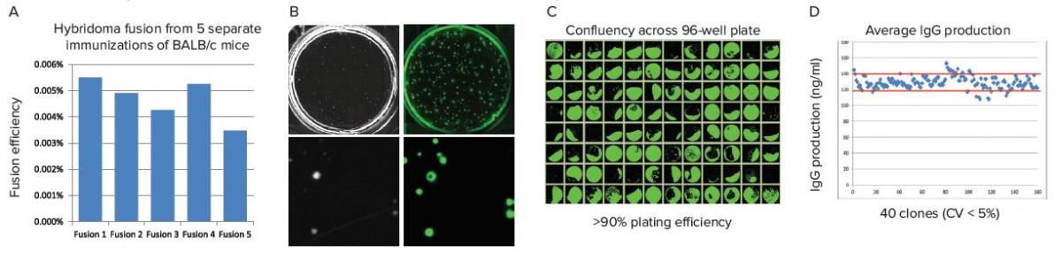
Figure 1:(A) Five individual hybridoma fusion experiments were conducted on BALB/c mice, immunized to the same antigen, to assess reproductibility of fusions in XP Media suite of products. Fusion efficiency was calculated by dividing the number of hybridoma colonies detected on the ClonePix 2 System by the number of splenocytes grown in XP Media Pre-Fusion Myeloma Growth Medium and Hybridoma Expansion Medium (without HT). (B) Images of hybridomas were captured with the ClonePix 2 System in white light (left panel) and FITC (right panel), after 7 days growth, to determine growth and expression of IgGs, respectively. Colonies grown in the presence of CloenDetect were ranked according to their FITC intensity (indicating total IgG production), with the highest producers picked for further characterization. (C) Software detection of cell confluency, indicated by the green overlay, across a 96-well plate allowing for a quick visualization of plating efficiency. 87 out of 96 wells grew to a confluency >5% after 7 days (the initial confluency of all wells was <1%) for a >90% plating effiency. (D) IgG production plotted per well (shown in blue) with red lines indicating 2 s.d. away from the mean. Because these are stable hybridomas, we don’t expect a large variation in the total amount of IgG produced per cell, which is confirmed by <5% CV across all clones tested.
Superior viability and growth of hybridomas in CloneMedia semi-solid media
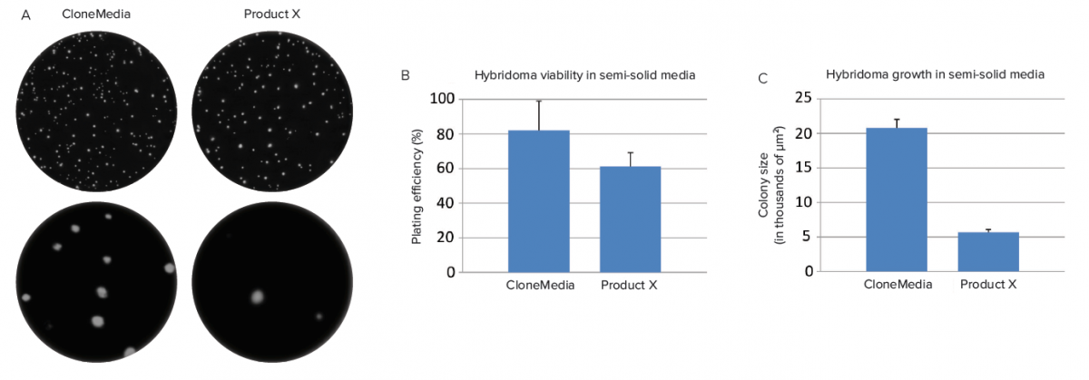
Figure 2:(A) Hybridomas were plated in CloneMedia semi-solid media from Molecular Devices and in semi-solid media from Competitor X. A greater number of clones were able to grow in CloneMedia when compared to Product X as shown in the white light images taken of colonies on the ClonePix 2 System. (B) Hybridoma viability was calculated by dividing the number of colonies detected by the initial seeding density. Hybridomas were more viable in CloneMedia than in Product X. (C) This was further confirmed by comparing the average colony size in both media.
Optimal growth and antibody production of hybridomas in liquid media
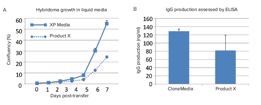
Figure 3:(A) Hybridomas grown in XP Media grew 2X as fast as Product X but did not suffer from a reduction in antibody production due to enhanced growth, as shown in (B).
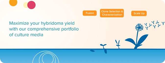
Workflow for antibody discovery in hybridomas
The traditional approach to generating monoclonal antibody-producing cells (i.e. hybridomas) is to fuse myeloma cells with desired antibody-producing splenocytes (e.g. B cells). These B cells are typically sourced from animals, usually mice. Following cell fusion, large numbers of clones are screened and selected on the basis of antigen specificity and immunoglobulin class. Once candidate cell line clones are identified, each “hit” is re-confirmed, validated, and characterized using a variety of downstream functional assays. Once completed, the clones are expanded or scaled up where additional downstream bioprocesses occur.
Here we provide a general overview of the hybridoma workflow showing where our XP Media and CloneMedia would be used in this hybridoma generation and scale-up process.
Download the hybridoma cell line development workflow >>

Related Collateral
Video
XP Media and CloneMedia for Mouse Hybridoma Generation
- Tutorial video: Hybridoma selection using HAT medium
- Tutorial video: Preparing and plating cells in semi-solid medium
Data Sheet
Poster
Brochures
eBook
User Guide
FAQ
| Product Name. | P/N | Format | Contains | Storage Temperature |
|---|---|---|---|---|
| XP Media and CloneMedia Complete Kit for Mouse Hybridoma Generation | K8861 | 1 Kit |
1 each of the following part numbers:
|
Please see below for storage of individual components |
| XP Media Pre-Fusion Myeloma Growth Medium and Hybridoma Expansion Medium (without HT) | K8862 | 500 mL |
|
|
| XP Media Hybridoma Fusion Medium | K8863 | 500 mL |
|
|
| XP Media Hybridoma Fusion Recovery Medium | K8864 | 100 mL |
|
|
| CloneMedia Hybridoma Semi-Solid Selection and Cloning Medium (with HAT) | K8865 | 90 mL |
|
|
| XP Media Hybridoma Growth Medium (with HT) | K8866 | 500 mL |
|
|
| CloneMedia Hybridoma Semi-Solid Selection and Cloning Medium (without HAT) | K8867 | 90 mL |
|
|
| Hybridoma Polyethylene Glycol (PEG) for Cell Fusion | K8868 | 1.5 mL |
|
|
*Heat-inactivated